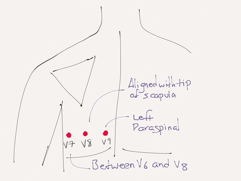
Where should a CCMA place the electrodes for leads V7 V8 and V9. V8 is placed at the tip of the left scapula in the same horizontal plane.

Electrocardiographic Diagnosis Of Remote Posterior Wall Myocardial Infarction Using Unipolar Posterior Lead V9 Chest
Divide 1500 by the number of small boxes between two R waves.

. Leads V7-9 are placed on the posterior chest wall in the following positions see diagram below. Level with V8 just left of vertebral line Special Lead Placement. Therefore in patients presenting with.
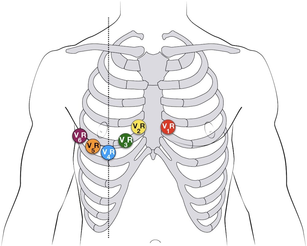
L V4 V5 V6 V7 V8 V9 AR V1 2 3 LL LL LL RA RA RA LA LA LA RL RL RL V1 V1 V1 V2 V2 LV2 V3 LV3RA V3 placement. Move V4 V5 V6 to posterior positions V7. V8 same horizontal line as V4R mid subscapular line use V5 electrode.
In none of the Group A or B patients was there ST elevation in leads V7 V8 or V9 either at rest or at peak exercise. V7 is located at the same horizontal line as V4R ie 5th ICS on the posterior axillary line use the V4 electrode. V8 is placed at the tip of the left scapula in the same horizontal plane.
On the posterior scapular line V9. On the left border of the spine. V8 Tip of the left scapula in the same horizontal plane as V6.
V1 V2 V3 V7 V8 and V9 are identical to the American ECGEKG. Level with V7 at mid-scapular line V9. Placement of Posterior Leads.
What is the correct placement of leads V7 V9. In the setting of the Acute Inferior STEMI the patient will frequently have an Acute Posterior and or RV STEMI. Leads I II and III.
This blog aims to disrupt how medical providers and trainees can gain public access to high-quality educational content while also engaging in a dialogue about best-practices in EM and medical education. If you make the diagnosis of Inferior STEMI you should routinely request leads V4R V7 V8 V9. Where are leads v7 v8 and v9 placed.
V9 is placed in the left paraspinal region in the same horizontal plane. Lead V7 V8 and V9 were recorded at the same horizontal level of lead V6 on the posterior axillary line lead V7 the posterior scapular line lead V8 and the left border. Inferior angle of the scapula.
V7 Left posterior axillary line in the same horizontal plane as V6. Basic 12-Lead Placement 1. Position trainer in the desired upright or horizontal position.
The initial ECG recorded the 12 classic leads and subsequently the 3 additional posterior leads V7. V8 Tip of the left scapula in the same horizontal plane as V6. Lead placement may vary by institution or instruction.
Placement of posterior leads V7-V9. Feel for anatomical landmarks on trainer remove electrode from sheet and place adhesive side. V7 is placed at the posterior axillary line in the same horizontal plane as V6.
V9 Left paraspinal region in the same horizontal plane as V6. An ECG lead is a graphical description of the electrical activity of the heart. Read full answer here.
From electrodes to limb leads chest leads 12-lead ECG. ST elevation in leads V7 V8 and V9 is uncommon in patients presenting with subendocardial ischaemia. Before discussing the ECG leads and various lead systems we need to clarify the difference between ECG leads and ECG electrodesAn electrode is a conductive pad that is attached to the skin and enables recording of electrical currents.
Posterior Ventricular leads V7 V8 V9. V9 same horizontal line as V4R left paraspinal border use V6 electrode. See figures 8 9 3.
Leads V7-9 are placed on the posterior chest wall in the following positions. When do you request these leads. When doing a right-sided EKG what is the placement of the leads.
Level with V6 at left posterior axillary line V8. These areas are most accurately monitored by the placement of special leads V4R V7 V8 V9. Basic 12-Lead Placement 1.
On the posterior axillary line V8. What are the lead groups that represents Einthovens Triangle. V7 Left posterior axillary line in the same horizontal plane as V6.
Lay out labeled leads and plug them into their designated outlets on the 15-lead electronics box. V9 is placed in the left paraspinal region in the same horizontal plane. ST depression was seen in 69 in V7 31 in V8 and 11 in V9 in the Group A patients at peak exercise.
Just to the lateral to the vertebrae. Ensure the trainer is clean. V7 is placed at the posterior axillary line in the same horizontal plane as V6.
V9 Left paraspinal region in the same horizontal plane as V6.

Active Chest Pain Trop 5 0 Core Im Podcast

Posterior Electrode Placement V7 Is Placed In The Left Posterior Download Scientific Diagram
Diagnostics Alternative Ekg Leads Taming The Sru

Ecg Lead Positioning Litfl Ecg Library Basics

Posterior Myocardial Infarction How Accurate Is The Flipped Ecg Trick
Lead Placement For Posterior Ecg Resus Review
Ecg Lead Positioning Litfl Ecg Library Basics

How To Not Miss A Posterior Myocardial Infarction Em Daily
0 comments
Post a Comment